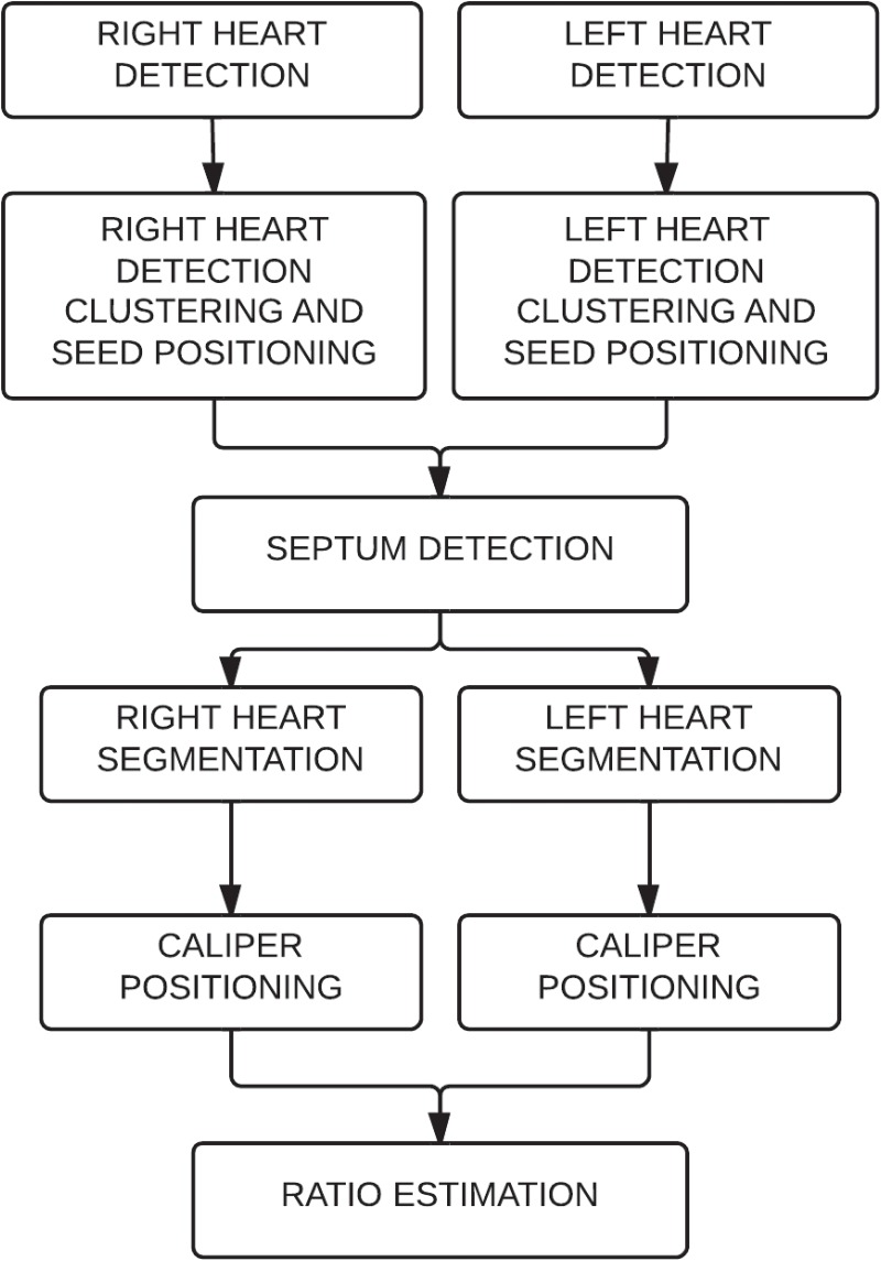
Fig 1. Algorithm description.
First, the right ventricle and the left ventricle are detected on each axial slice. The detections are clustered to position seeds for further segmentation. The seeds are used to detect the septum. Using the seeds and the septum, the ventricles are segmented and the calipers positioned. Finally the right ventricular to left ventricular axial diameter ratio is estimated.

