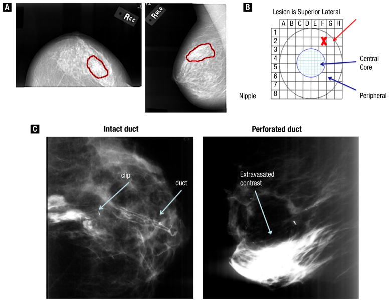
Figure 2.
Ductal Carcinoma In Situ Duct (DCIS) Identification. (A) Technique for Locating Nipple Opening That Corresponds to the Area on Mammogram. (B) Grid That Represents the Nipple Surface. (C) Ductogram, Showing Contrast Agent Localized to Within the Duct (left) or Leaking From the Perforated Duct into the Breast and Stroma (right)
MRI = magnetic resonance imaging; PLD = pegylated liposomal doxorubicin.

