
Figure 6. Testing the Lin B TF code: dendritic arborization.
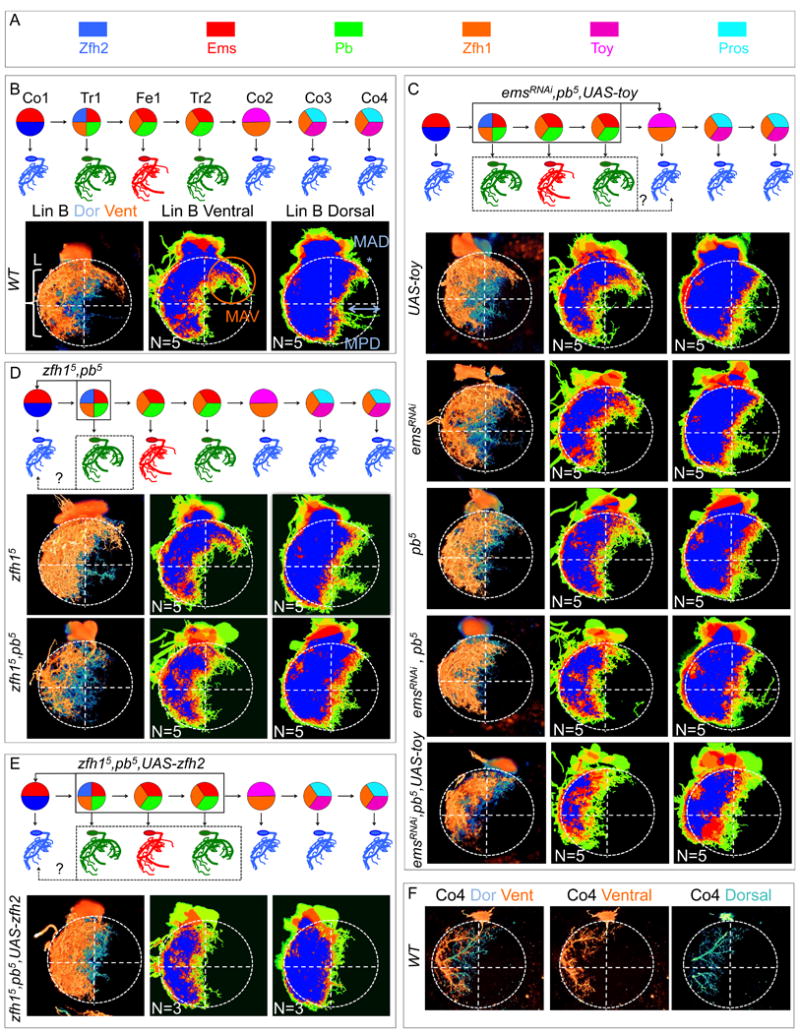
(A): TF color key.
(B): Top, birth order, TF code, and schematic of dendritic arbors for the seven wild type Lin B MNs. Bottom left, WT Lin B MARCM clone in the T1 hemisegment of an adult VNC in which the ventral (orange) and dorsal (blue) hemispheres are pseudo-colored; bottom right, heat maps of ventral and dorsal hemispheres illustrating the degree of overlap for five independent samples.
(C): From top to bottom: schematic of expected transformation in emsRNAi, pb-/-, UAS-toy MARCM clones; UAS-toy; emsRNAi; pb-/-; emsRNAi, pb-/- and emsRNAi, pb-/-, UAS-toy Lin B MARCM clones.
(D): From top to bottom: schematic of expected transformation in zfh1-/-, pb-/- MARCM clones; zfh1-/- and zfh1-/-, pb-/- Lin B MARCM clones.
(E): Top: schematic of expected transformation in zfh1-/-, pb-/-, UAS-zfh2 Lin B MARCM clones; bottom: zfh1-/-, pb-/-, UAS-zfh2 Lin B MARCM clone.
(F): WT Co4 MN labeled with MARCMbow in which the ventral (orange) and dorsal (blue) hemispheres are pseudo-colored. Note the similarity to the arborization pattern at bottom of panels C, D and E.