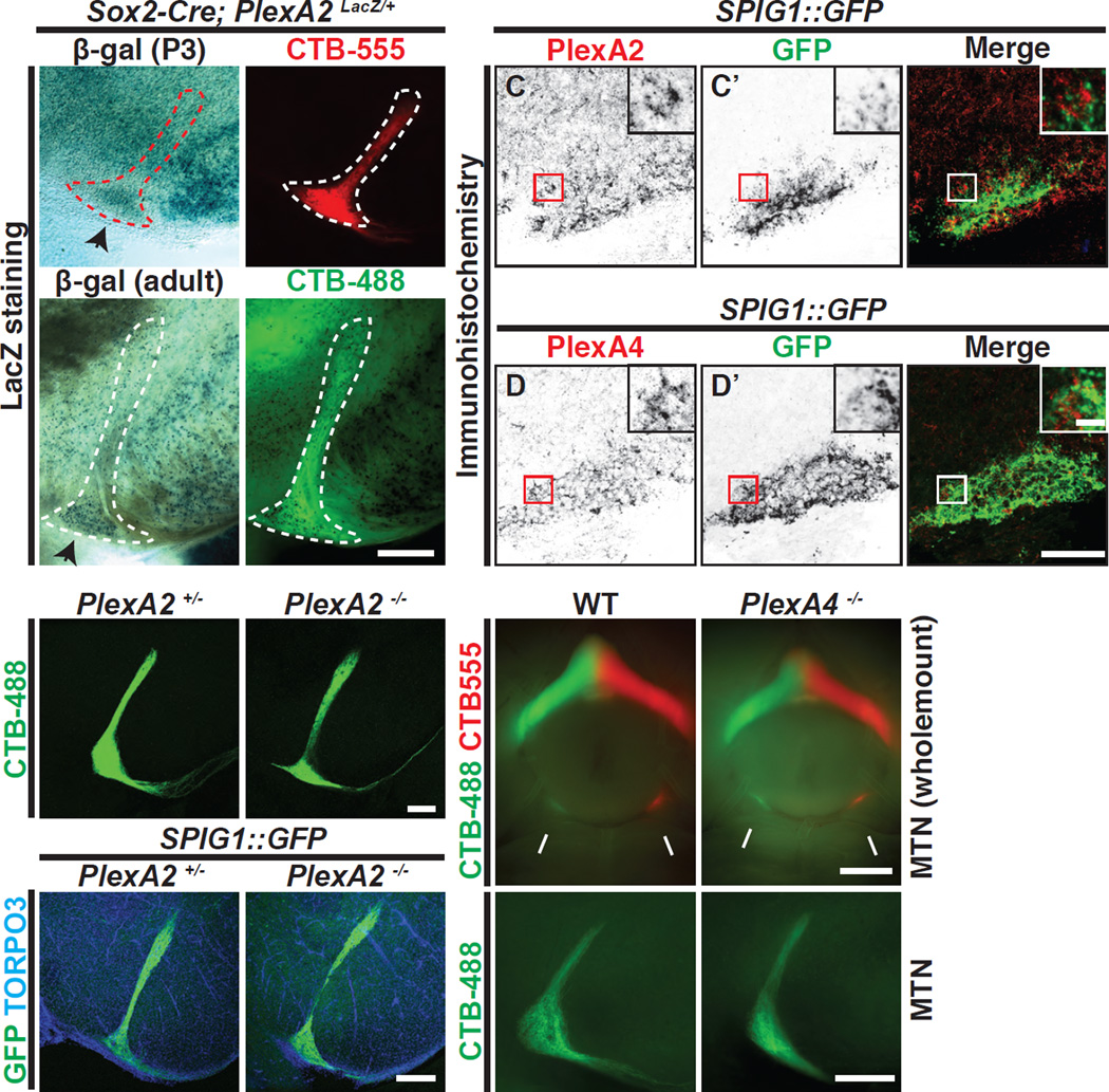
Figure 4. PlexA2 and A4 are Expressed in the MTN, and PlexA2−/− and PlexA4−/− Single Mutants Exhibit Normal On DSGC-MTN Innervation.
(A-B’) Expression of PlexA2 in the MTN using ocular CTB injections (A’ and B’) and the PlexA2 LacZ reporter encoded in the PlexA2 conditional allele (Sun et al., 2013) (A and B), revealing that PlexA2LacZ+ cells are abundant in the ventral region of the MTN at P3 and in the adult (black arrowheads in A and B, respectively; n=3 animals at each developmental stage). (CD”) Immunostaining of PlexA2 (C) and PlexA4 proteins (D) in the MTN at e18.5 along with SPIG1::GFP+ axon fibers to delineate the ventral MTN region (C’ and D’). PlexA2 and PlexA4 proteins (C and D, respectively) are expressed in the MTN region, and PlexA2+ and PlexA4+ cells (red boxes in C and D, respectively) are in very close proximity to SPIG1::GFP+ fibers (red boxes in C’ and D’, and white boxes in C” and D”). (E–H) Characterization of RGC-MTN innervation in PlexA2+/− (E and G) and PlexA2−/− (F and H) animals by ocular CTB injections (E and F) and by genetic labeling (G and H). No obvious defects are observed in PlexA2−/− mutants (n=6 PlexA2+/− mice, n=8 PlexA2−/− mice; n=3 SPIG1::GFP; PlexA2+/− and 3 SPIG1::GFP; PlexA2−/− mice). (I–L) Wholemount ventral view (I and J) and cross-sectional view (K and L) of On-DSGC MTN innervation visualized by ocular CTB injections in WT (I and K) and PlexA4−/− mutants (J and L). On DSGC-MTN innervation appears normal in PlexA4−/− mutants (compare I and J, K and L; also see (Matsuoka et al., 2011b) Supplementary Figure 6) (n=4 WT; n=5 PlexA4−/− mutants). Scale bars: 250 µm in (B’) for (A)-(B’); 100 µm in (D”) for (C)-(D”); 10 µm in the inset of (D”) for insets of (C)-(D”); 250 µm in (F) for (E) and (F); 250 µm in (H) for (G) and (H); 1 mm in (J) for (I) and (J); and 250 µm in (L) for (K) and (L).

