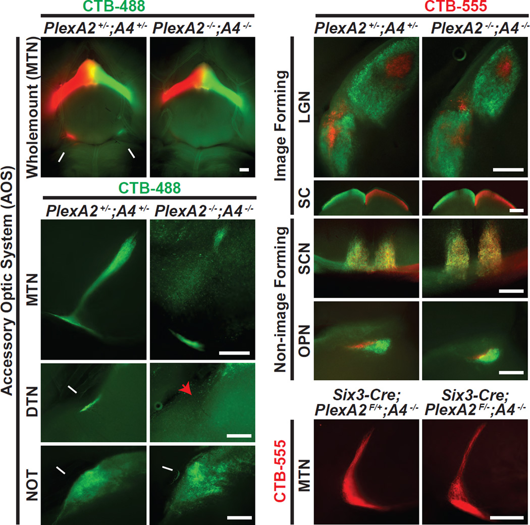
Figure 5. PlexA2−/−;PlexA4−/− Double Mutants Phenocopy Sema6A−/− Single Mutants with Respect to On DSGC-MTN Innervation.
(A and A’) Ventral wholemount view of PlexA2+/−;PlexA4+/− (A) and PlexA2−/−;PlexA4−/− (A’) adult brains following ocular injections with CTB-488 and CTB-555 bilaterally. Prominent projections to the MTN were observed in PlexA2+/−;PlexA4+/− animals (white arrows in A), whereas MTN innervation is greatly diminished in PlexA2−/−;PlexA4−/− mutants (A’), phenocopying Sema6A−/− mutants (n=9 PlexA2+/−; PlexA4+/− mice; n=9 PlexA2−/−; PlexA4−/− mutants, with 8/9 showing full expressivity). (B-D’) Cross-sectional views of the accessory optic visual system in PlexA2+/−;PlexA4+/− (B–D) and PlexA2−/−;PlexA4−/− (B’-D’) mice. Compared to controls, PlexA2−/−;PlexA4−/− mutants exhibit greatly reduced MTN innervation (B’), a complete loss of DTN innervation (red arrowhead in C’), but apparently normal NOT innervation (white arrow in D’). (E-H’) Characterization of RGC axonal innervation to image-forming (E, E’, F, F’) and non-image-forming (G, G’, H, H’) retinorecipient targets in controls (E–H) and PlexA2−/−;PlexA4−/− mutants (E’-H’). In comparison to controls, the segregation of ipsilateral and contralateral retinal projections and general retinorecipient targeting are preserved in PlexA2−/−;PlexA4−/− mutants (n=9 PlexA2+/−;PlexA4+/− mice; n=9 PlexA2−/−;PlexA4−/− mutants). (I and I’) Removal of PlexA2 genetically in a PlexA4−/− mutant background (I’) results in similar MTN innervation as in the control (I), showing that retina-derived PlexA2 is not required for normal development of On DSGC-MTN projections (n=4 animals for both genotypes). Scale bars: 250 µm.

