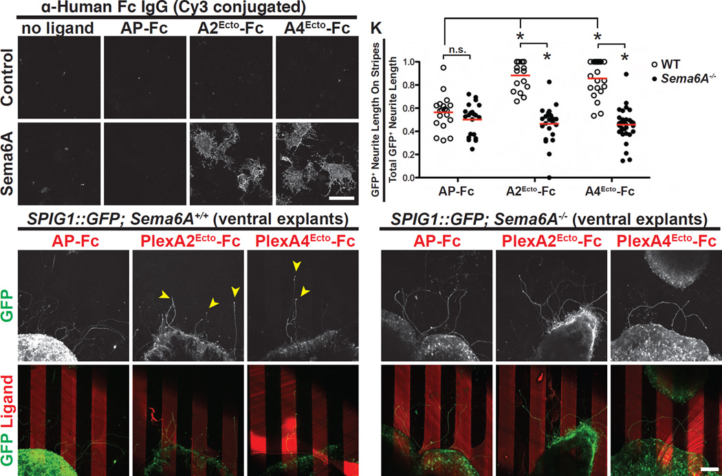
Figure 6. PlexA2 and A4 Function as Attractive Cues for SPIG1::GFP+ On DSGC Neurites in vitro.
(A-D’) Fluorescent ligand binding assay showing that PlexA2 and PlexA4 ectodomain recombinant proteins only bind to COS7 cells that express mouse Sema6A in vitro (C’ and D’). (E-G’) The ventral region of SPIG1::GFP; Sema6A+/+ retinas was dissected out and cultured on plates coated with recombinant proteins presented as “stripes” (Sun et al., 2013). After two days in vitro, SPIG1::GFP+ neurites that extended from explants show no preferences for AP-Fc stripes (E and E’). In contrast, SPIG1::GFP+ neurites preferentially grew on PlexA2Ecto-Fc and PlexA4Ecto-Fc recombinant protein stripes (yellow arrowheads in F and G, respectively) (n=19 explants on AP-Fc stripes, n=17 explants on PlexA2ecto-Fc stripes, and n=21 explants on PlexA4ecto-Fc stripes; the explants were from 9 embryos). (H-J’) SPIG1::GFP+ neurites extending from SPIG1::GFP; Sema6A−/− ventral retina explants exhibit no preference for AP-Fc (H and H’), PlexA2Ecto-Fc (I and I’), or PlexA4Ecto-Fc stripes (J and J’) (n=21 explants on AP-Fc stripes, n=24 explants on PlexA2Ecto-Fc stripes, and n=35 explants on PlexA4Ecto-Fc stripes; explants were from 9 embryos). (K) Quantification of GFP+ neurite length on stripes over the total GFP+ neurite length in the stripe assay. Red bars represent the mean for each group. *P<10−6. Scale bars: 100 µm in (D’) for (A)-(D’); and 100 µm in (J’) for (E)-(J’).

