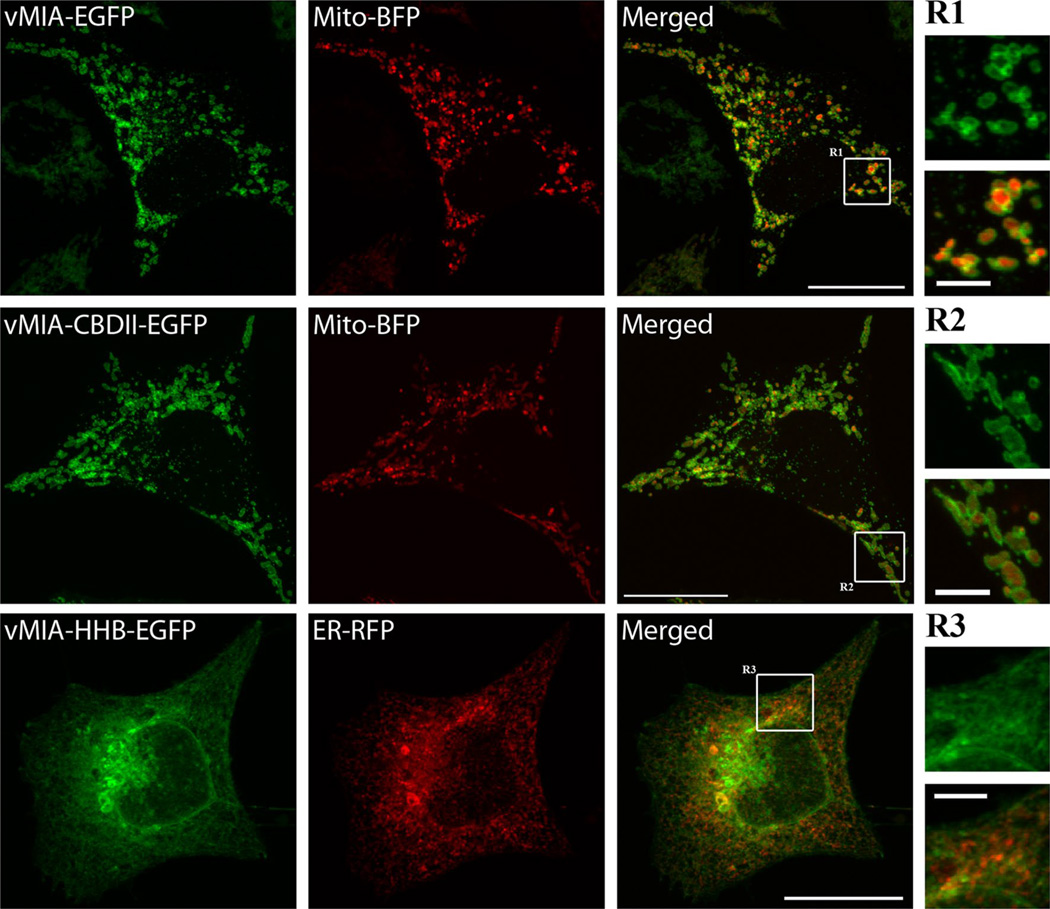
Fig. 1.
Sub-cellular localization of vMIA wild type, and vMIA-CBDII, and vMIA-HHB mutants. HeLa cells were transiently transfected with plasmid vectors expressing vMIA-enhanced green fluorescent protein (EGFP), vMIA-CBDII-EGFP and vMIA-HHB-EGFP using Lipofectamine 2000 as previously described [93]. Plasmids expressing ER (ER-red fluorescent protein, RFP fused with KDEL) and mitochondrial (Mito-blue fluorescent protein, BFP) markers were used for co-transfection. Cells were fixed with 4 % paraformaldehyde for 15 min 20–24 h after transfection. Fixed cells were imaged using an Olympus FV1000 confocal microscope. Images shown are a single plane of vMIA-EGFP (green) and Mito-BFP (pseudocolored red) transfected HeLa cells (top row), vMIA-CBDII-EGFP (green) and Mito-BFP (pseudocolored red) transfected HeLa cells (middle row) and vMIA-HHB-EGFP (green) and ER-RFP (red) transfected cells (bottom row). Zooms of the regions R 1, 2, and 3 show the distribution of vMIA-EGFP (R1), vMIA-CBDII (R2), and vMIA-HHB (R3) in green (top) and merged (bottom) panels. The bars on the merged full-size images represent 20 µm and on the zoomed regions represent 4 µm

