Fig. 1.

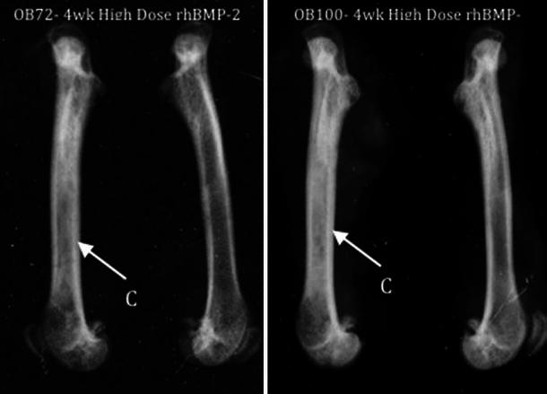
Magnified lateral radiographs of re-sected femora at 4 weeks. C Qualitative observation of cortical thickening in rhBMP-2 treated femurs. Note: treated (right) femurs appear on the left in both radiographic images

Magnified lateral radiographs of re-sected femora at 4 weeks. C Qualitative observation of cortical thickening in rhBMP-2 treated femurs. Note: treated (right) femurs appear on the left in both radiographic images