Fig. 2.

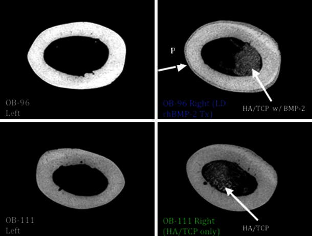
MicroCT midshaft cortical scan: note new periosteal bone growth (P) in the LD rhBMP-2 treated femur on the top right compared to contralateral limb and HA/TCP-only treated sample

MicroCT midshaft cortical scan: note new periosteal bone growth (P) in the LD rhBMP-2 treated femur on the top right compared to contralateral limb and HA/TCP-only treated sample