Fig. 3.

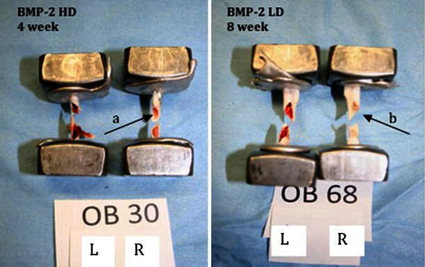
Spiral fractures resulting from torsional testing. Pictures show mid-diaphyseal spiral fractures. Note that the IM canals of the treated right femora appear lighterred (a) and white (b) due to the presence of HA/TCP carrier

Spiral fractures resulting from torsional testing. Pictures show mid-diaphyseal spiral fractures. Note that the IM canals of the treated right femora appear lighterred (a) and white (b) due to the presence of HA/TCP carrier