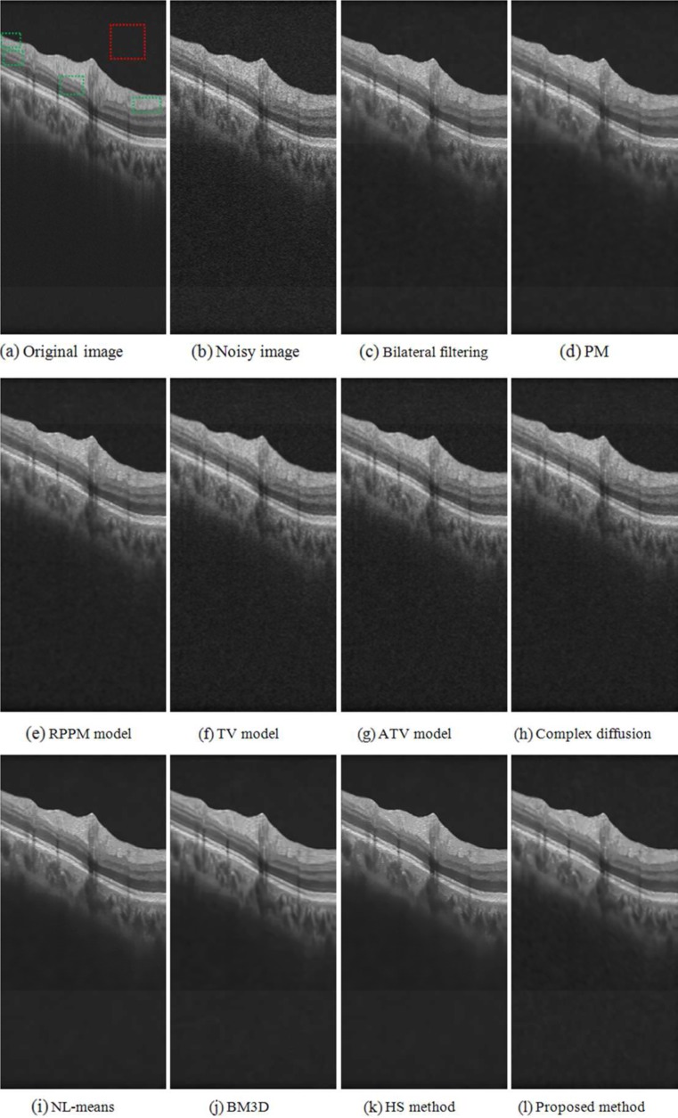
Fig. 8.
Image denoising of a synthetic noisy OCT retinal image. a Original “noise-free” B-scan. The red square indicates an example location identified as background region, the green rectangles indicate example locations considered as foreground regions. b B-scan corrupted by synthetic noise. c to i Results obtained from the different denoised methods investigated

