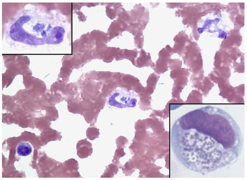
Figure 2.

Anaplasma phagocytophilum-infected band neutrophil in human peripheral blood (Wright stain, original magnification × 260). The top left inset shows the same band neutrophil and demonstrates several morulae with a stippled basophilic appearance corresponding to individual bacteria (magnification × 520). The bottom right insert shows A. phagocytophilum cultivated in vitro in the human promyelocytic leukemia cell line HL-60. Here, the individual basophilic bacteria are easily visualized within vacuoles of the infected cell (LeukoStat stain; magnification × 520).
