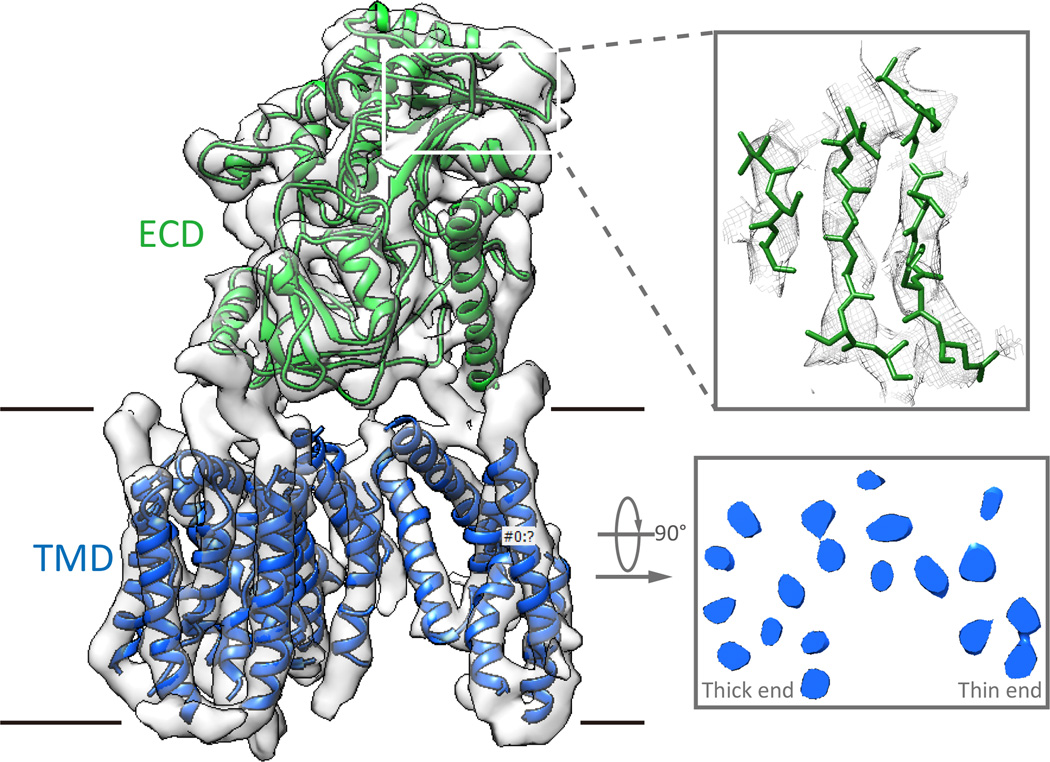
Figure 5. Near-atomic resolution cryo-EM structure of human gamma-secretase.
(A) Overall view of the complex with the trans-membrane domain (TMD), which is made up of the four different proteins, in blue, and the extra-cellular domain (ECD) of Nicastrin in green. (B) Representative density for the soluble domain showing a region of the map with separated betastrands. (C) View inside the TMD perpendicular to the membrane showing the horse-shoe like arrangement of the trans-membrane helices with a thick and a thin end. The lack of good sidechain density in this region of the map prohibited the assignment of each helix to the four different proteins.

