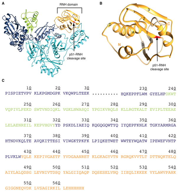
Figure 1.
Ribbon representation of the structures of (A) p66/p51 RT heterodimer and (B) the RNH domain, indicating the p51-RNH processing site (arrow), and (C) amino acid sequence of p66. In (A)-(C), the Thumb and RNH domains in the p66 subunit are shown in green and orange, respectively; the p51 subunit in cyan. Structures in (A) and (B) were drawn using PDB:1DLO.

