Figure 6.

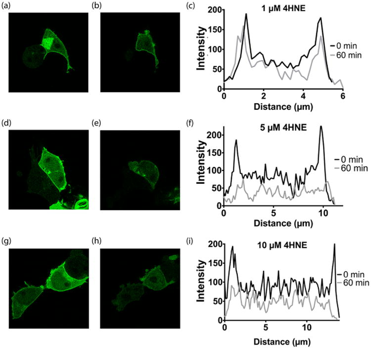
Subcellular localization of RGS4 during 4HNE treatment. RGS4 interaction with its native binding partner, Gαo, was evaluated by monitoring subcellular localization of GFP-RGS4 in HEK293T. (a) Prior to treatment with 1 μM 4HNE, GFP-RGS4 strongly associates with the plasma membrane. (b) After 1 h treatment with 1 μM 4HNE, GFP-RGS4 showed no change in localization. (c) Quantification of GFP-RGS4 fluorescence in a cross-section of both pretreatment (a) and post-treatment with 4HNE (b). Prior to treatment, the pixel intensity of GFP-RGS4 at the plasma membrane was 280% of the cell body. After 1 h of treatment with 1 μM 4HNE, the pixel intensity ratio of GFP-RGS4 at the plasma membrane as compared to the cell body remained unchanged. (d) Prior to treatment with 5 μM 4HNE, GFP-RGS4 strongly associates with the plasma membrane. (e) After 1 h treatment with 5 μM 4HNE, GFP-RGS4 localization to the plasma membrane is completely reversed. (f) Quantification of GFP-RGS4 fluorescence in a cross-section of both pretreatment (d) and post-treatment with 4HNE (e). Prior to treatment, the pixel intensity of GFP-RGS4 at the plasma membrane was 260% of the cell body. After 1 h of treatment with 5 μM 4HNE, the pixel intensity of GFP-RGS4 at the plasma membrane fell to within 50% above the mean intensity of the plasma membrane. (g) Prior to treatment with 10 μM 4HNE, GFP-RGS4 strongly associates with the plasma membrane. (h) After 1 h treatment with 10 μM 4HNE, GFP-RGS4 localization to the plasma membrane is completely reversed. (i) Quantification of GFP-RGS4 fluorescence in a cross-section of both pretreatment (g) and post-treatment with 4HNE (h). Prior to treatment, the pixel intensity of GFP-RGS4 at the plasma membrane was 250% of the cell body. After 1 h of treatment with 10 μM 4HNE, the pixel intensity of GFP-RGS4 at the plasma membrane fell to within 50% above the mean intensity of the plasma membrane. The images shown are representative of n = 3 experiments.
