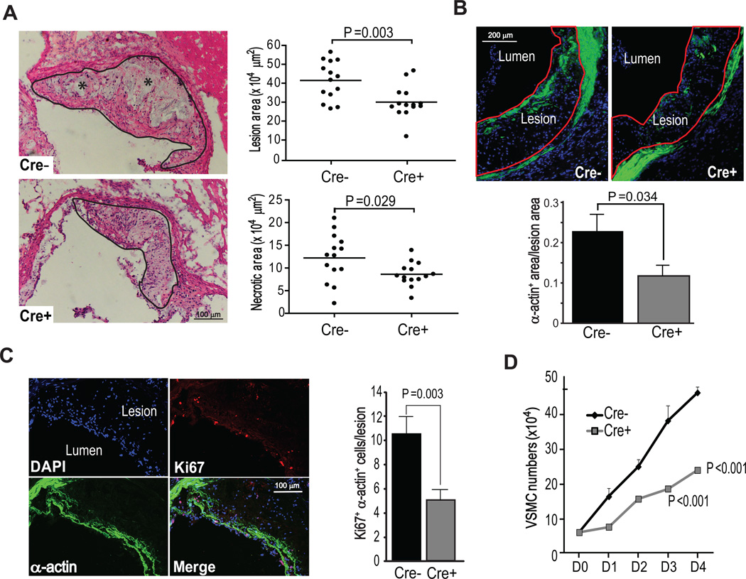
Figure 1. CHOP deletion in VSMCs in Apoe−/− mice reduces the content of α-actin-positive cells in atherosclerotic lesions and suppresses VSMC proliferation.
A, Representative images and quantitative analyses of aortic root lesion area and necrotic area in Chopfl/flApoe−/− and Chopfl/flSM22α-CreKI+Apoe−/− mice (n=14). The lesions are delineated with a black line, and necrotic areas are shown by asterisks. B, Representative images of α-actin staining (green) and quantification of proportion of the α-actin-positive area in Chopfl/flApoe−/− and Chopfl/flSM22α-CreKI+Apoe−/− aortic root lesions (n=8). The lesions are delineated with a red line. C, Representative Z series projection images of Ki67 (red) and α-actin staining (green) in Chopfl/flApoe−/− lesions and quantification of Ki67/α-actin-positive VSMCs (n=8). The upper left image shows the DAPI+ cells in this projection. Scale bar, 50 µm. D, Proliferation curve of Chopfl/flApoe−/− and Chopfl/flSM22α-CreKI+Apoe−/− VSMCs cultured in 10% FBS medium 0–4 days (D0–D4) after serum-starvation (n=3).

