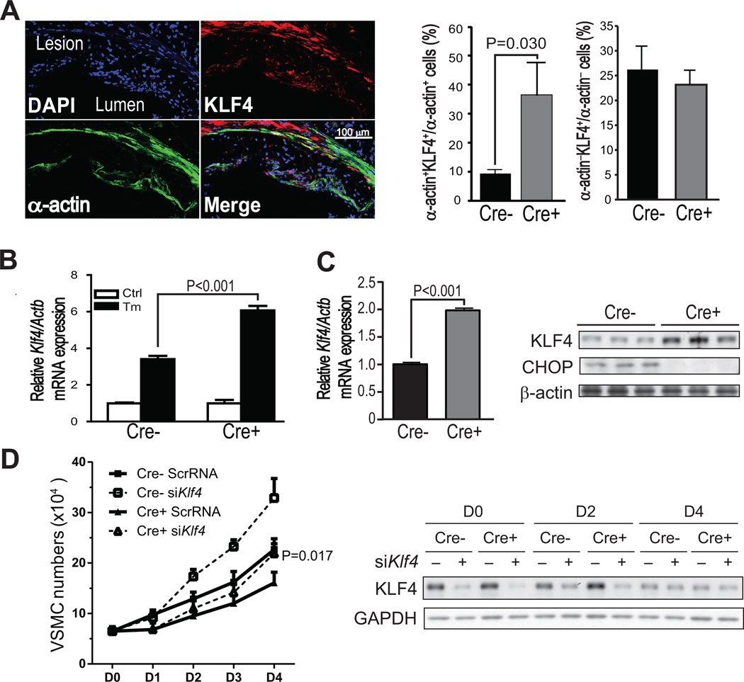
Figure 2. KLF4 is increased in α-actin-positive cells in Chopfl/flSM22α-CreKI+Apoe−/− lesions and is a mechanism of decreased VSMC proliferation.
A, Representative Z series projection images of KLF4 staining (red) and α-actin staining (green) in Chopfl/flSM22α-CreKI+Apoe−/− lesions and quantification of KLF4+;α-actin+ cells or KLF4+;α-actin− cells in Chopfl/flApoe−/− and Chopfl/flSM22α-CreKI+Apoe−/− lesions (n=8). The upper left image shows the DAPI+ cells in this projection. Scale bar, 100 µm. B, Klf4 mRNA relative to Actb in Chopfl/flApoe−/− and Chopfl/flSM22α-CreKI+Apoe−/− VSMCs treated with 2.5 µg/ml tunicamycin for 12 h (n=3). C, Klf4 mRNA relative to Actb (n=6, graph) and immunoblot of KLF4 and CHOP protein (n=3) in Chopfl/flApoe−/− and Chopfl/flSM22α-CreKI+Apoe−/− VSMCs cultured in 10% FBS medium for 24 hours after serum starvation. β-actin was included as loading control. D, Left panel: Proliferation curve of scrambled (Scr) or Klf4 siRNA-transfected Chopfl/flApoe−/− and Chopfl/flSM22α-CreKI+Apoe−/− VSMCs cultured in 10% FBS medium 0–4 days after starvation (n=3; at day 4, the Cre+ siKlf4 value is different from the Cre+ ScrRNA value at P=0.017). Right panel: KLF4 protein expression in Scr or Klf4 siRNA-transfected Chopfl/flApoe−/− and Chopfl/flSM22α-CreKI+Apoe−/− VSMCs.

