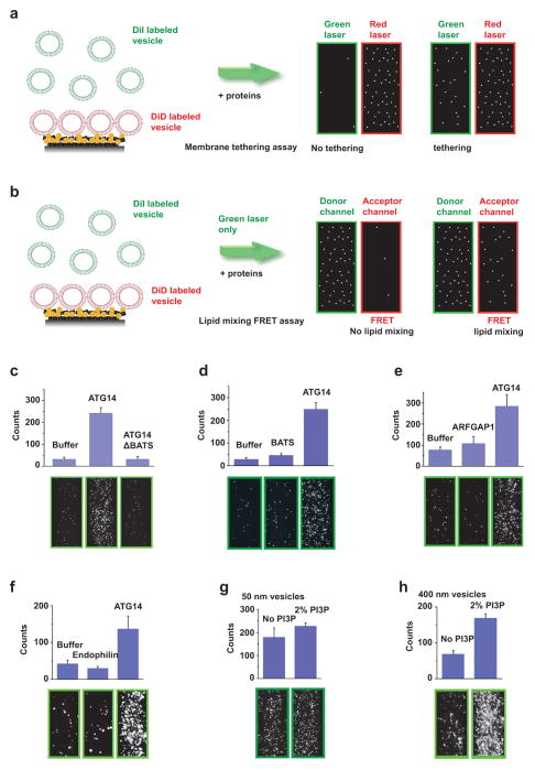
Extended Data Figure 3. Characterization of ATG14-mediated membrane tethering.
a, Scheme of the single-vesicle/liposome-tethering assay13,14. DiD-labelled liposomes were attached to the imaging surface through the interaction between biotin/NeutrAvidin; surface binding was assessed by red laser excitation (633 nm). Tethered DiI-labelled liposomes were detected by green laser excitation (532 nm). This assay was used in Figs 2a and 4e and Extended Data Figs 3c–h, 5d and 5g. b, Scheme of the FRET-based single-vesicle/liposome lipid-mixing assay13. Tethered DiI-labelled liposomes were excited by illumination with a green laser (532 nm). Detection of emission in both green and red spectral regions was performed simultaneously by using a dichroic beam-splitter. The total number of tethered DiI-labelled liposomes were counted in the green fluorescence channel, and FRET to the DiD acceptor dyes was observed in the red fluorescence channel. This assay was only used in Fig. 2b. The field of view is 45 μm ×90 μm. See Methods for more details. c, The BATS domain deletion mutant of ATG14 (900 nM) does not promote liposome tethering. Top panels: quantitation. Bottom panels: representative fluorescence images of tethered liposomes (n =15). d, The BATS domain alone does not promote liposome tethering. Top panels: quantitation. Bottom panels: representative fluorescence images of tethered liposomes (n =15). e, Purified recombinant ADP-ribosylation factor GTPase-activating protein 1 (ARFGAP1, 870 nM) does not promote liposome tethering. Top panels: quantitation. Bottom panels: representative fluorescence images of tethered liposomes (n =15). f, Purified recombinant endophilin 1/Bif 1 (2 μM) does not promote liposome tethering. Top panels: quantitation. Bottom panels: representative fluorescence images of tethered liposomes (n =15). g, Incorporation of 2% PI3P into small liposomes (with a 50 nm diameter) failed to enhance the liposome-tethering activity by ATG14. Top panels: quantitation. Bottom panels: representative fluorescence images of tethered liposomes (n =15). h, Incorporation of 2% PI3P into large liposomes (with a 400 nm diameter) enhances the liposome-tethering activity by ATG14. Top panels: quantitation. Bottom panels: representative fluorescence images of tethered liposomes (n =15). Results in c–h are presented as the mean (±s.d.) of random imaging locations (n =15) in the same sample channel.

