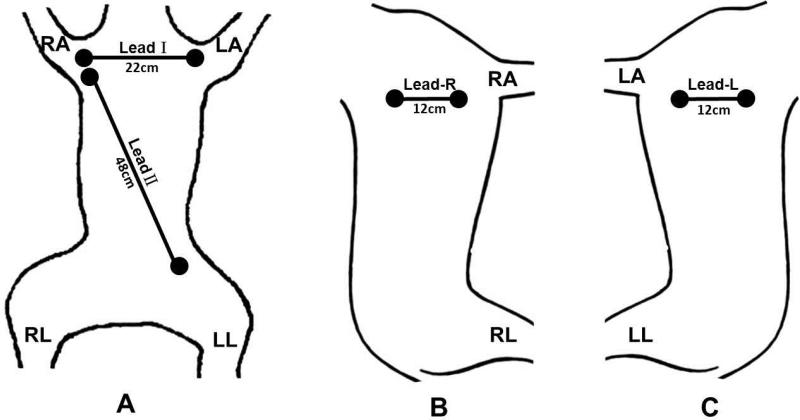
Figure 1.
Schematics of electrode location on the surface of the skin showing dogs in supine (A), left decubitus (B) and right decubitus (C) positions. In A, the two electrodes for Lead I recording were placed on the second rib and were 22 cm apart (11 cm from the midline). The Lead II electrodes were separated by 48 cm. B shows the location of the right bipolar electrode pair for recording SKNA-R. C shows the location of the left bipolar electrode pair for recording SKNA-L.

