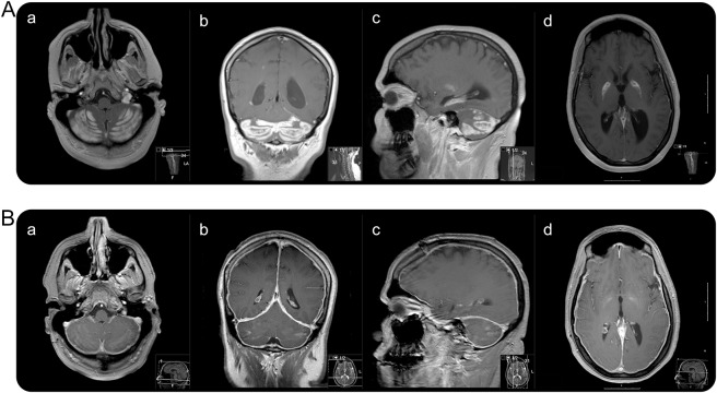
Figure 1. MRI at admission and 3-month follow-up.
(A) Initial T1-weighted imaging with contrast: axial (a), coronal (b), and sagittal (c) show cerebellar involvement. Axial (d) demonstrates globi pallidi involvement. (B) Three-month follow-up T1-weighted imaging with contrast: axial (a), coronal (b), and sagittal (c) show interval resolution of contrast enhancement. Axial (d) demonstrates resolution of globi pallidi enhancement. Note diffuse pachymeningeal enhancement.

