
FIGURE 5.
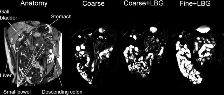
On the left side of the panel is a coronal anatomic “roadmap” dual-echo MRI image. The other images are corresponding small bowel water images taken across the small bowel of the same healthy young adult at time t = 4 h after consumption of each of the 3 meals containing a 20% sunflower oil in water emulsion on the 3 separate study days: Coarse, Coarse+LBG, and Fine+LBG. These images show the large amount of freely mobile fluid present in the small bowel in response to the fat emulsion meals. Coarse, 20% oil and water emulsion with 6-μm mean droplet size; Coarse+LBG, 20% oil and water emulsion with 6-μm mean droplet size and 0.5% locust bean gum; Fine+LBG, 20% oil and water emulsion with 0.4-μm mean droplet size and 0.5% locust bean gum; LBG, locust bean gum.