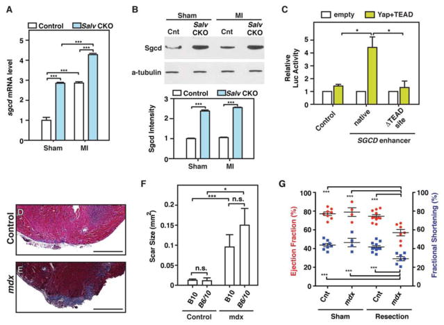
Figure 6. The dystrophic complex is downstream of the Hippo pathway and is required for cardiac regeneration.
(A, B) LADO and sham surgery were performed in control and Salv CKO mouse hearts at P8, and heart samples were collected at 4 dpmi. Delta-sarcoglycan (Sgcd) mRNA was detected in control and Salv CKO mouse hearts by using qRT-PCR and was normalized to Gapdh (N=3 hearts for all groups) (A). Sgcd protein was detected by using Western blot analysis (N=3 hearts per genotype and treatment) (B). Sgcd band intensities were quantified and normalized to those of alpha-tubulin. ***P<0.001, remaining column comparisons were non-significant (n.s.) (C) Luciferase assays were performed with P19 embryonal carcinoma cells. Cells were transfected with either the control luciferase reporter, a reporter containing the Sgcd enhancer, or a reporter containing the Sgcd enhancer but lacking the Tead site. Three independent experiments with technical triplicates were performed. *P<0.05. (D–G) Dystophin glycoprotein complex (DGC) is required for endogenous cardiac regeneration. Representative images of trichrome-stained heart sections from B10 control (D) and Mdx-B10 (E) mice subjected to resection of the cardiac apex. Images of 2 additional control and mutant apexes are shown in fig. S9. Bars=500 μm. Quantification (F) of the scar size at 21 dpr in B10, (N=11), B6/10 (N=4), Mdx-B10 (N=7), and Mdx-B6/10 (N=6) mouse hearts.*P<0.05, ***P<0.001. Echocardiography analysis (G) of control sham (N=3), control apex resection (n=7), Mdx sham (N=4), and Mdx apex resection (N=7) mouse hearts 21 days after surgery. ***P<0.001, remaining column comparisons were n.s.

