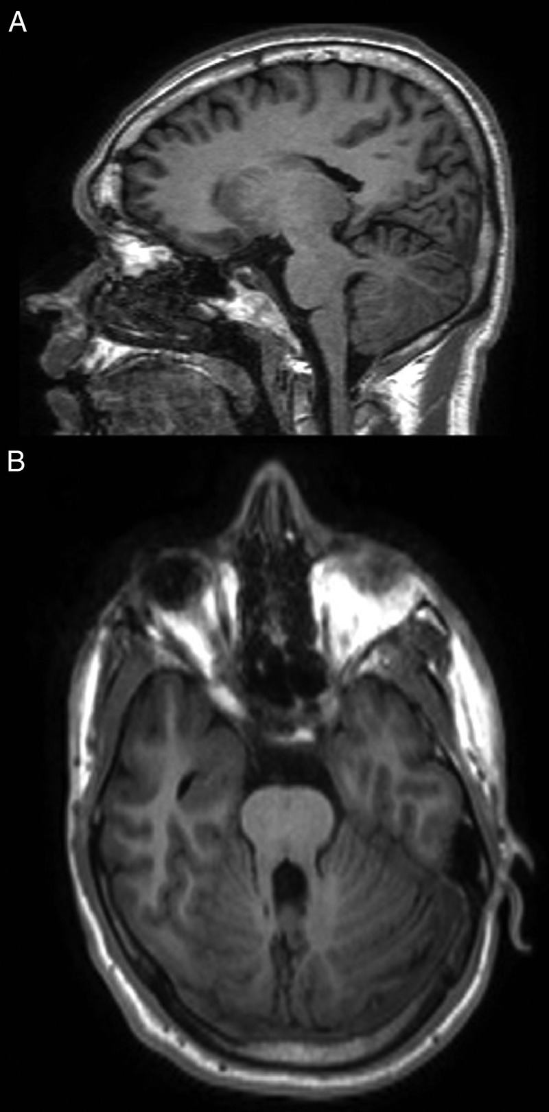
Figure 1.

(A) Sagittal MRI T1-weighted images through the cerebellum and brain stem of the patient, depicting the horizontalisation of the superior cerebellar peduncles and other hindbrain anomalies. (B) Note the ‘molar tooth sign’ on the axial plane.
