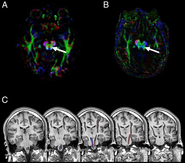
Figure 3.

(A) Diffusion tensor imaging (DTI)-tractography showing normal decussation of the corticospinal tract as a red dot between the cerebral pyramids (arrow). (B) DTI-tractography of our patient, showing the absence of decussation. Colours represent tract orientation in space; red, green and blue represent the x, y and z-axis, respectively. (C) DTI-tractography over T1-weighted sequential coronal planes of the brainstem, illustrating lack of decussation at the medullar pyramids.
