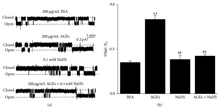
Figure 1.
AGEs-induced activation of ENaC is reversed by 0.1 mM NaHS in A6 cells. (a) The representative ENaC single-channel current recorded from A6 cells, respectively, treated with basolateral 200 μg/mL BSA (control; top trace), basolateral 200 μg/mL AGEs, apical 0.1 mM NaHS, and basolateral 200 μg/mL AGEs + apical 0.1 mM NaHS (bottom trace) for 24 h. (b) Summary plot shows that AGEs treatment significantly increased ENaC P O, which was reversed by H2S treatment (n = 10 for each individual experimental set; ∗∗ indicates P < 0.01 compared to control; ## indicates P < 0.01 compared to AGEs treated cells).

