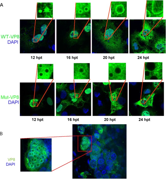
FIG 6.
Cellular localization of WT-VP8 and Mut-VP8. (A) Localization and different patterns of WT-VP8 and Mut-VP8 in the nucleus. WT-VP8 and Mut-VP8 were expressed in COS-7 cells. VP8 was detected with polyclonal anti-VP8 antibody and Alexa-488-conjugated goat anti-rabbit IgG. DNA was labeled with DAPI. The cells were observed with a Zeiss LSM410 confocal microscope. (B) Localization of VP8 in BoHV-1-infected lung tissue slices. Lung tissue sections (220 to 250 μm) were infected with 106 PFU of BoHV-1 for 24 h. VP8 was detected with polyclonal anti-VP8 antibody and Alexa-488-conjugated goat anti-rabbit IgG. DNA was labeled with DAPI. The slides were observed with a Zeiss LSM410 confocal microscope.

