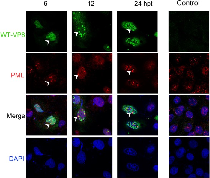
FIG 7.
PML protein accumulation to nuclear bodies and colocalization with WT-VP8. (A) pFLAG-VP8 was transfected into COS-7 cells. FLAG-WT-VP8 was detected with monoclonal anti-FLAG antibody and Alexa-488-conjugated goat anti-mouse IgG, and PML protein was detected with polyclonal anti-PML antibody and Alexa-633-conjugated goat anti-rabbit IgG. DNA was labeled with DAPI. The cells were observed with a Zeiss LSM410 confocal microscope. The white arrowheads indicate that at 6 hpt PML protein is recruited to the edge of nuclear bodies, where WT-VP8 accumulates, and that at 24 hpt nuclear PML accumulates to the bodies, resulting in protein clusters.

