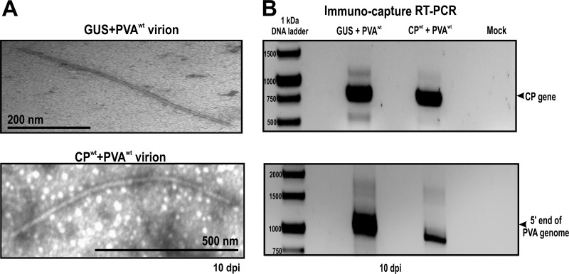
FIG 7.
Transmission electron microscopy (TEM) and immunocapture RT-PCR. Virion formation was assessed in Nicotiana benthamiana plants coexpressing PVAwt with GUS (control plants) or CPwt (experimental plants) at 10 dpi. (A) Virions contained in both control and experimental plants were captured on carbon-coated grids, followed by negative staining with 3% uranyl acetate for visualization on a JEOL 1400 TEM. Virions similar in shape and size were detected in both plants. (B) Immunocapture RT-PCR was used to investigate the RNA content of the virus particles. Control and experimental plants both contained the full-length RNA molecule.

