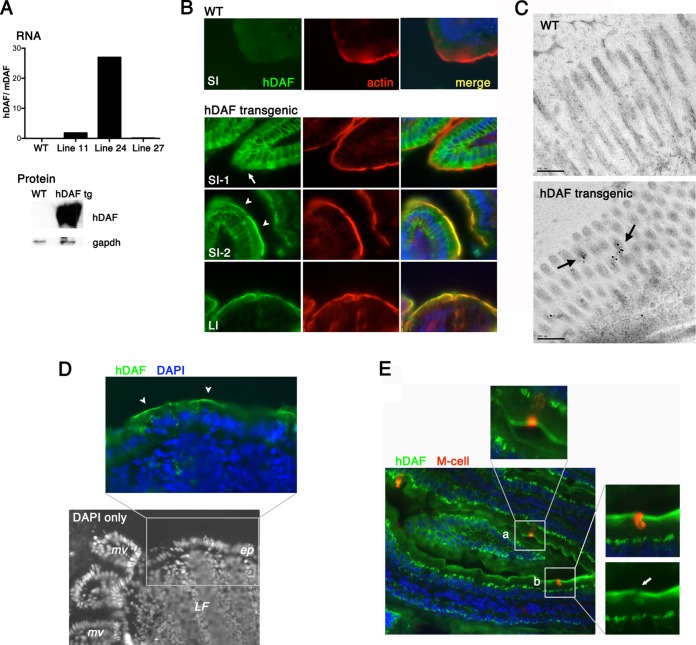
FIG 1.
hDAF expressed on the epithelium of transgenic mice. (A) hDAF RNA and protein expression. (Top) RNA was measured by real-time PCR to assess hDAF RNA expression in the intestines of wild-type (WT) and transgenic mice (lines 11, 24, and 27). Data are shown as the ratio of the hDAF RNA copy number to the copy number for endogenous murine DAF RNA. (Bottom) Immunoblot to measure hDAF protein expression in intestines of wild-type and transgenic (tg) animals (line 24). (B) Immunofluorescence detection of hDAF in the intestines of wild-type and transgenic animals. In the small intestine (SI), hDAF (green) was detected both within cells (SI-1, arrow) and on the apical cell surface (SI-2, arrowheads), marked by phalloidin staining of the cortical actin layer (red). In the large intestine (LI), hDAF was expressed on the apical cell surface. (C) Immunogold electron microcopy to detect DAF in the small intestine. Gold label (arrows) is associated with the microvilli in transgenic but not wild-type mice. (D) Black-and-white image of DAPI-stained distal small intestine, showing a lymphoid follicle (LF) with overlying epithelium (ep) and adjacent microvilli (mv). The expanded color panel shows follicle-associated epithelium stained for hDAF (green); arrowheads indicate DAF at the apical epithelial cell surface. (E) Small intestine stained for hDAF (green) and an M cell marker (red). Some M cells appear to stain for surface DAF (inset a); however, the red M cell staining is associated with a neighboring cell in a different focal plane (inset b, arrow).

