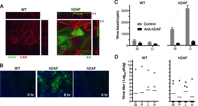
FIG 3.
Human DAF permits infection of primary murine intestinal cells in vitro, but hDAF transgenic mice resist infection by the oral route, despite virus attachment to hDAF on intestinal epithelium. (A) Primary intestinal epithelial cells from hDAF transgenic or wild-type (WT) mice were cultured as polarized monolayers on Transwell filters and stained with anti-DAF (green) and anti-CAR (red) antibodies, as described in Materials and Methods. DAF is present on the apical surface of monolayers from hDAF but not WT mice. (B) Polarized monolayers were exposed to CVB3 H3-RD (5 PFU per cell) for 1 h at 16°C, and then monolayers were washed and incubated at 37°C to permit infection to proceed. At 8 h, monolayers were stained to detect newly synthesized viral capsid protein VP1; input virus (0 h) was not detectable. (C) Intestinal segments (1 cm long) were treated with medium containing anti-hDAF MAb IF7 or with medium alone (Control) and then exposed to 35S-labeled CVB3 H3-RD, and binding was determined as described in Materials and Methods. Results are indicated as the mean cpm bound + SD for triplicate samples. (D) Wild-type mice (n = 4) and hDAF transgenic mice (n = 5) were exposed to 1 × 10⁹ PFU of CVB3 H3-RD by gavage, and virus titers in tissues were determined at 3 days postinfection. SI, small intestine; P, pancreas; L, liver; H, heart; LI, large intestine.

