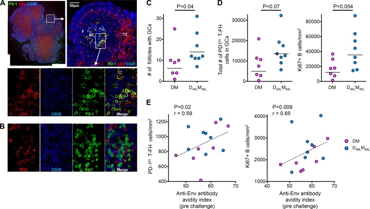
FIG 3.
D40LM40L vaccine enhances T-FH and germinal center B cell responses. (A) Immunofluorescence imaging of a nondraining lymph node from a CD40L-adjuvanted animal at 2 weeks after the final MVA boost shows germinal center follicles with T-FH cells. Sections were stained with CD4 (red), CD20 (blue), and PD-1 (green) as described previously (7, 8). The merged image shows PD-1high CD4 T-FH cells abundantly present within germinal centers of lymphoid follicles. F, follicle; GC, germinal center; TZ, T cell zone. Scale bars = 50 microns. (B) Sections of lymph node germinal center follicles stained with CD3 (red), CD20 (blue), and Ki67 (green) show proliferating (Ki67+) B cells in germinal center follicles. (C) Numbers of follicles with germinal centers in nonadjuvanted (DM) and adjuvanted (D40LM40L) animals. The P value reflects significance as determined by the Mann-Whitney t test. (D) Total number of T-FH cells and density of Ki67+ B cells/mm2 of section. The total number of T-FH cells was calculated as a product of the density of T-FH cells/mm2 and the number of follicles in the section. (E) Correlation of the density of T-FH cells and Ki67+ B cells with the avidity index of anti-SIVmac239 Env IgG at the time of challenge. P values reflect significance as determined by Spearman's rank correlation test. These analyses were performed on seven DM and eight D40LM40L animals.

