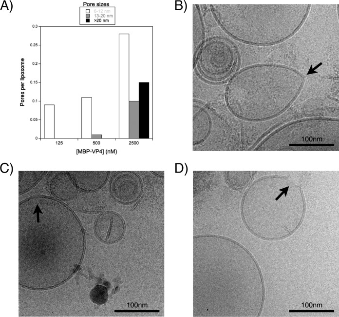
FIG 6.
Cryo-EM imaging. (A) Pore sizes observed by cryo-EM. PA-PC (1:1 molar ratio) liposomes at 500 μM were incubated with different protein concentrations. Membrane interruptions observed in digital micrographs acquired at different nominal magnifications ranging from ×40,000 to ×80,000 were clustered in three different groups: 6 to 12 nm, 13 to 20 nm, and >20 nm. (B to D) Cryo-EM micrographs of membrane disruption. Interruptions in the membrane (black arrows) correspond to the groups with pore sizes of 6 to 12 nm (B), 13 to 20 nm (C) and >20 nm (D).

