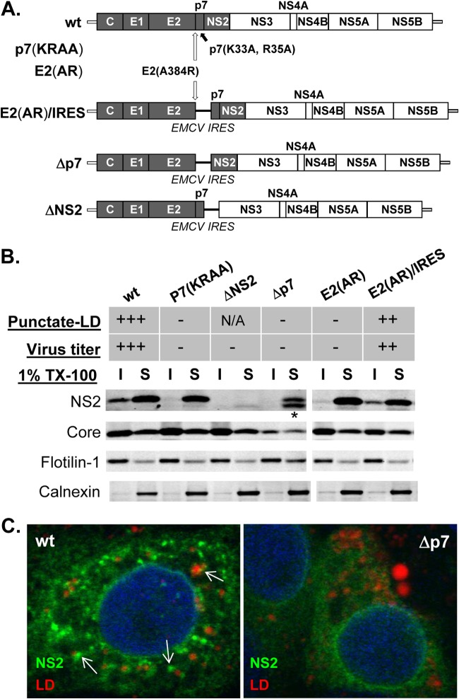
FIG 1.
E2-p7 processing and p7-dependent NS2 subcellular localization to the detergent-insoluble fraction (DIF) in HCV-replicating cells. (A) Organization of wild-type (wt) HCV and various HCV mutants used in this study. Note that the H77 sequence is shaded within this chimeric HCV encoding gt1a H77 Core to NS2 within the gt2a JFH1 background. (B) Western blots of cold 1% Triton X-100 (1% TX-100) lysates separated into the detergent-soluble (S) and -insoluble (I) fractions. NS2 localization to the punctate foci near lipid droplets (LD) as described before (10) is indicated in the Punctate-LD row as follows: +++, ∼80% of NS2-positive cells with this phenotype; ++, ∼50% of NS2-positive cells with this phenotype; −, background level; N/A, not available. Virus titers were >2 × 10E5 FFU/ml (+++), >2 × 10E4 (++), and undetectable (−). The asterisk at the bottom of NS2 from Δp7 indicates the presence of additional bands detectable by NS2 antibody from this mutant and may represent an aberrant NS2 initiation product by EMCV IRES as described previously (16). (C) Confocal image analysis by using an Zeiss LSM 510 Meta laser-scanning confocal microscope of cells at day 2 postelectroporation with HCV RNA encoding the indicated genomes. Anti-NS2 antibody (green) and LipidTOX deep red neutral lipid stain (red) were used to detect NS2 and lipid droplets. Examples of the punctate-LD phenotype of NS2 are indicated by the white arrows.

