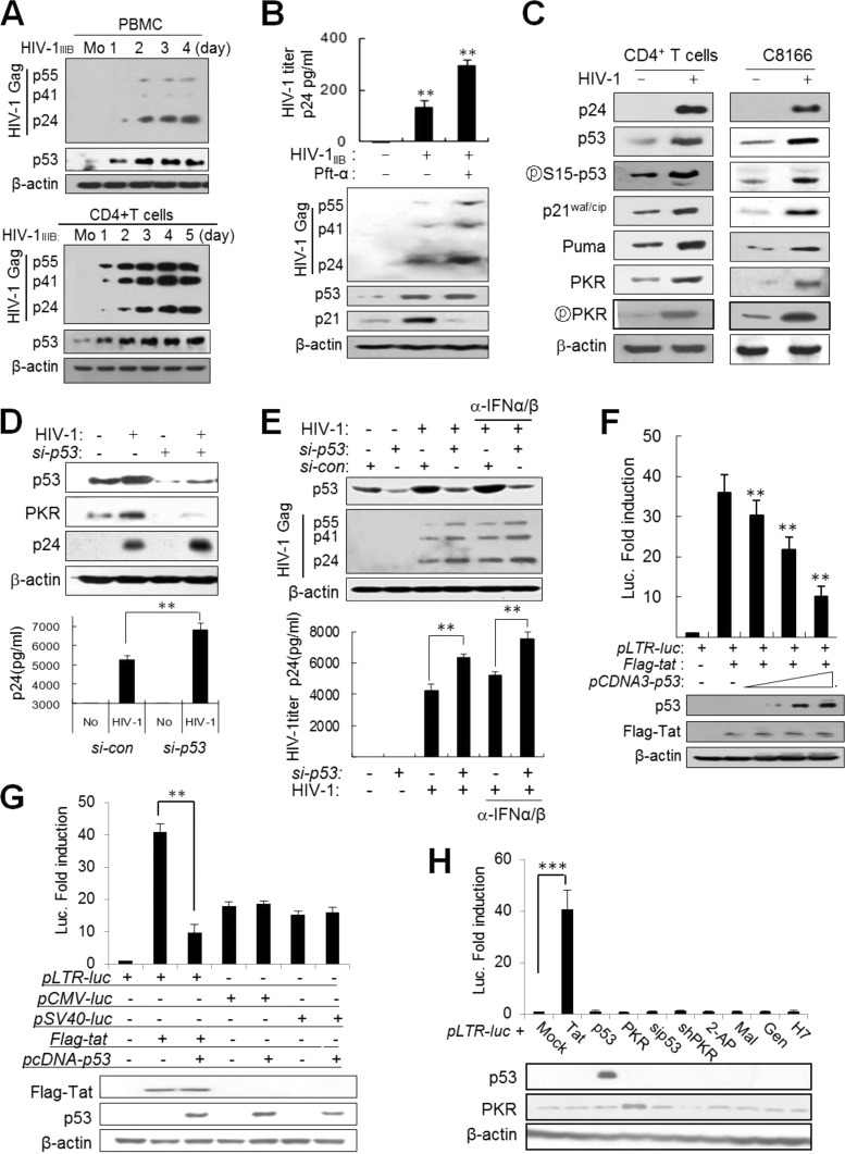
FIG 1.
HIV-1 infection induces the expression of p53, and p53 inhibits HIV-1 replication by suppression of Tat activity through PKR. (A) Preactivated PBMCs (upper panel) and CD4+ T cells (lower panel) were infected with HIV-1IIIB at an MOI of 1 for the indicated periods of time, and the levels of p53 and HIV-1 Gag proteins in infected cells were assessed by Western blotting with anti-p53 and anti-p24 antibodies. Mo, mock infection. (B) Upper panel, HIV-1-infected PBMCs were cultured in the presence or absence of 10 μM pifithrin-α (Pft-α) (a p53 inhibitor). Four days after infection, titers of virus in culture supernatants were assessed by p24 ELISA and are represented as means ± SD (n = 3). **, P < 0.01. Lower panel, HIV-1 Gag proteins in infected cells were assessed by Western blotting. (C) The expression of p53 and p53 target genes was assessed 4 days after infection of CD4+ T cells with HIV-1IIIB and 40 h postinfection of C8166 cells with HIV-1 (HXBc2) at an MOI of 1. (D) HIV-1 replication was assessed in p53-normal and p53KD (si-p53) (p53 siRNA; Cell Signaling) C8166 cells 48 h after infection (MOI of 1) from cell extracts by Western blotting (upper panel) and from culture supernatants using a p24 ELISA kit (represented as means ± SD; n = 3) (lower panel). **, P < 0.01 for untreated versus treated cells. (E) Effects of p53 and type 1 interferon (IFN) on HIV-1 replication and PKR expression. Upper panel, p53-normal (si-con) and p53KD (si-p53) C8166 cells were infected with HIV-1 (HXB2) at an MOI of 1 in the presence or absence of 5 μg/ml anti-IFN-α/β neutralizing antibodies (BioLegend Inc.). After 48 h, cell extracts were assessed by Western blotting with anti-HIV-1 Gag antibodies. Lower panel, the titers of HIV-1 in culture supernatants were assessed in triplicates at 48 h postinfection using a p24 ELISA kit (Perkin-Elmer) and are represented as means ± SD (n = 3). **, P < 0.01 versus control siRNA (si-con). (F) HCT116 p53−/− cells were transfected with pLTR-luc (200 ng), pcDNA-Flag-tat (100 ng), and increasing amounts of pcDNA3-p53 (1, 2.5, and 5 μg), together with 20 ng of pCMV-lacZ as a transfection control. Luciferase activity was assessed 2 days after transfection and expressed after normalization to β-galactosidase activity. Data are represented as means ± SD (n = 4). Protein levels were assessed by Western blotting after normalization to β-actin. **, P < 0.01. (G) pLTR-luc, pCMV-luc, and pSV40-luc reporter plasmids were cotransfected with the indicated Tat or p53 plasmid combinations into HeLa cells together with pLTR-luc as a transfection control. Luciferase activity was expressed after normalization with the β-galactosidase activity of each reaction. Protein levels were assessed by Western blotting after normalization with β-actin. **, P < 0.01. (H) HeLa cells were cotransfected with pLTR-luc and other expression vectors together with pCMV-lacZ as a transfection control. In addition, HeLa cells transfected with pLTR-luc and p pCMV-lacZ were treated with each kinase inhibitor as shown. Luciferase activity was assessed. ***, P < 0.001.

