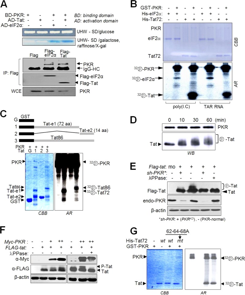
FIG 3.
PKR phosphorylates the first exon of HIV-1 Tat. (A) Upper panel, yeast two-hybrid experiments with PKR and Tat as described in Materials and Methods. Lower panel, endogenous PKR was assessed by Co-IP with anti-Flag Ab in C8166 cells transfected with pcDNA-Flag-tat/eIF2α. (B) Recombinant GST-PKR protein (0.5 μg), preactivated with poly(I·C) or TAR RNA for 1 h, was incubated with 0.5 μg of recombinant His-eIF2α or His-Tat proteins in the presence of 1 μCi of [γ-32P]ATP, and phosphorylation of Tat and eIF2α was examined by autoradiography after SDS-PAGE. (C) Recombinant GST-Tat proteins encoding the first exon (72 aa), second exon (14 aa), or full-length Tat (86 aa) were incubated with preactivated PKR in the presence of [γ-32P]ATP, separated on a 12% polyacrylamide gel, and then assessed by autoradiography. (D) Preactivated PKR (GST-PKR) was incubated with column-purified His-Tat72 for 10 to 60 min at 30°C. Reaction mixtures were separated by 15% SDS-PAGE and then subjected to Western blot analysis. (E) PKRKD (sh-PKR+) and PKR-normal (−) HEK293 cells were transfected with pcDNA3-Flag-tat. Cells were treated with 50 nM calyculin A (a phosphatase inhibitor) for 1 h prior to harvest, and then cell extracts were assessed by Western blotting. Parts of samples were additionally treated for 1 h with λPPase (1 U/μl) before SDS-PAGE. (F) PKR-mediated Tat phosphorylation in vivo. HEK293 cells were cotransfected with pCDNA-Flag-tat and different concentrations of pCDNA-Myc-PKR. Two days after transfection, cells were treated with 50 nM calyculin A for 1 h prior to harvest, and cell extracts were separated by 15% SDS-PAGE and then subjected to Western blot analysis with anti-Flag, anti-Myc, and anti-β-actin antibodies. One of the samples was treated with λPPase prior to the Western blot assay. (G) wt or 62-64-68A mt Tat (72 aa) was incubated with preactivated PKR in the presence of [γ-32P]ATP. The reaction products were separated on a 12% SDS-polyacrylamide gel and then examined by autoradiography.

