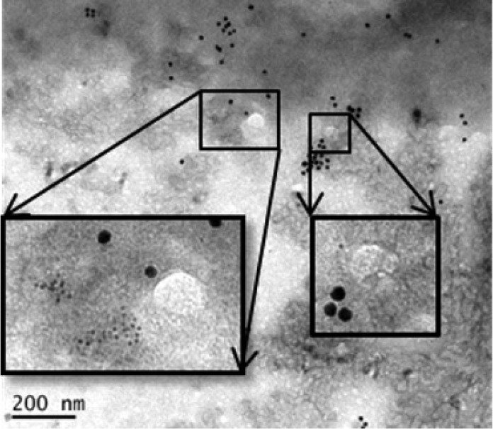
FIG 7.
Detection of NCL, SCARB2, and EV71 in RD cells. We stained EV71-infected RD cells with gold-labeled SCARB2 antibody (3-nm beads) and gold-labeled NCL antibody (13-nm beads). The whole-mount cells were viewed after a negative-staining procedure, followed by conventional transmission electron microscopy. EV71 particles are shown in white circles. Small black dots, SCARB2; large black dots, nucleolin. Insets are higher-magnification images.

