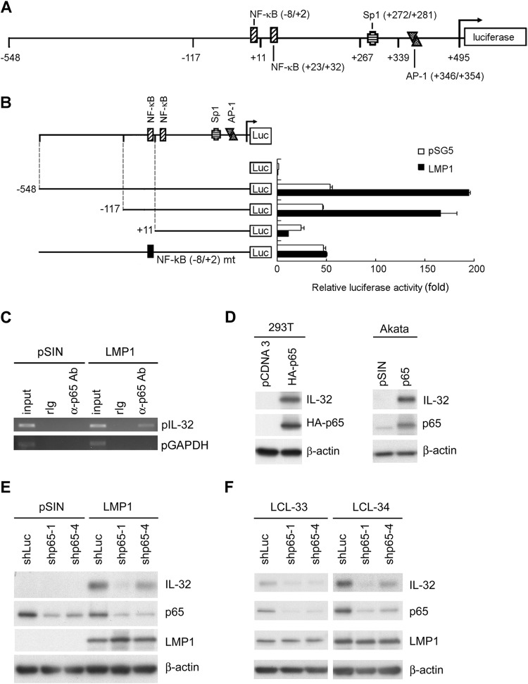
FIG 4.
LMP1 regulates IL-32 expression through p65 activation. (A) Schematic illustration of the reporter plasmids of the IL-32 promoter. Predicted binding sites of transcription factors on the promoter region are indicated. (B) 293T cells were transfected with LMP1-expressing plasmids, 5′ deletion IL-32 reporter plasmids, and pEGFP-C1 as a transfection control. After 72 h, the relative luciferase activity of each transfectant was normalized to its GFP intensity and standardized to the vector control cells. (C) Akata cells were infected with pSIN- and LMP1-expressing lentiviruses at an MOI of 1 for 5 days. Complexes of DNA and p65 were immunoprecipitated from the cells using anti-p65 antibody or rabbit IgG. DNA of IL-32 promoters (pIL-32) and GAPDH promoters (pGAPDH) were detected in the immunoprecipitates by PCR. Total DNA was harvested from vector- or LMP1-expressing cells and used as the input control. (D) 293T cells were transfected with p65-expressing plasmids for 3 days, and Akata cells were infected with p65 lentiviruses at an MOI of 1 for 5 days. Expression of IL-32, p65, and β-actin was measured by Western blotting. β-Actin served as the internal control. (E) Akata cells were coinfected with pSIN- or LMP1-expressing lentiviruses at an MOI of 1 and shLuciferase, shp65-1, or shp65-4 lentiviruses at an MOI of 4 for 5 days. Expression of IL-32, p65, LMP1, and β-actin was measured by Western blotting. β-Actin served as the internal control. (F) LCLs were infected with shLuciferase, shp65-1, or shp65-4 lentiviruses at an MOI of 4 for 5 days, and infected cells were further selected with 2 μg/ml of puromycin for another 2 days. Expression of IL-32, p65, LMP1, and β-actin was measured by Western blotting. β-Actin served as the internal control.

