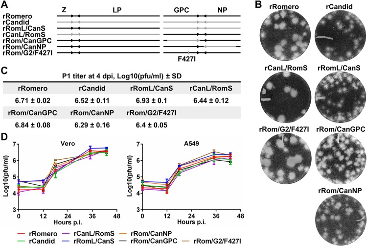
FIG 1.
Rescue and characterization of rJUNV with inter- and intrasegment rearrangements between the Rom and Can strains of JUNV. (A) Schematic representation of rJUNV genomes. Black and gray colors indicate the genetic material of Rom and Can strains of JUNV, respectively. (B) Plaque phenotype on Vero cells. Infected cell monolayers were overlaid with growth medium containing 0.5% agarose and incubated for 7 days. Cells were stained with crystal violet to visualize plaques. (C) Passage 1 (P1) titers on Vero cells were determined by plaque assay at 4 days p.i. (dpi). (D) One-step growth curves of rJUNVs on Vero (left) and A549 (right) cells. TCS of cells infected at an MOI of 5 were collected at the indicated times, and titers were determined by plaque assay.

