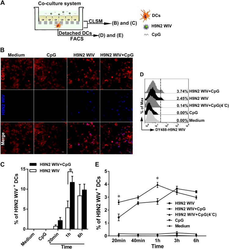
FIG 3.
Enhancement of H9N2 WIV uptake by submucosal DCs after CpG addition in the DC/EC coculture system. (A) Schematic of experimental setting to study the viral uptake in the coculture system. DCs were seeded on the underside of the filter facing the basolateral membrane of ECs for 4 h, and then, medium, H9N2 WIV (50 μg/ml HA), and/or CpGs (10 μg/ml) were incubated on the apical side of ECs. (B) Uptake of H9N2 WIV by submucosal DCs was determined using immunofluorescence. After 1 h, the filters in the coculture system were processed and views from between the DCs and the filter were obtained by CLSM. CD11c DCs (red) attached to the underside of the filter. DyLight 405-labeled H9N2 WIV (blue) existed within the submucosal DCs. Bars, 10 μm. (C) Quantification of virus-loaded submucosal DCs from fluorescence images. Values were calculated from six random fields of view (0.044 mm2 per field) for each of three individual filters. (D and E) FACS analysis of virus-loaded submucosal DCs. DCs were collected from the coculture system after 1 h (D) or after the indicated times (E) and detected by FACS. The data shown are the mean results ± SD from three independent experiments. *, P < 0.05.

