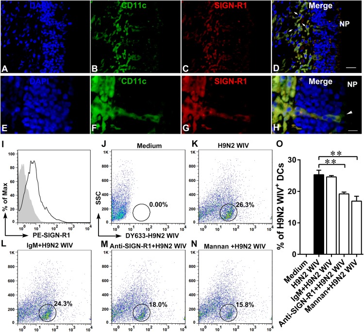
FIG 7.
Expression of SIGN-R1 receptor on DCs is conducive to H9N2 WIV uptake. Mice (n = 5) were nasally administered H9N2 WIV plus CpGs for 0.5 h, and then noses were collected. (A to H) CLSM analysis of SIGN-R1 receptor located in lamina propria DCs (A to D) or in TEDs (E to H). Blue, DAPI; green, CD11c; red, SIGN-R1; yellow, merge; arrows, lamina propria DCs; arrowheads, TEDs. Bars: 20 μm (A to D); 10 μm (E to H). (I) Histogram showing the results of FACS analysis of SIGN-R1 expression on DCs in vitro. Filled gray, isotype control; black line, stained cells. (J to O) In vitro, DCs were pretreated with or without SIGN-R1-specific blocking antibody (20 μg/ml) or isotype IgM (20 μg/ml) or mannan (1 mg/ml) for 30 min at 37°C and then incubated with DyLight 633-labeled H9N2 WIV (HA concentration of 10 μg/ml) for 30 min. (J to N) FACS analyses of viral uptake by DCs. SSC, side scatter. (O) Quantification of the FACS results as shown in panels J to N. Data shown are the mean results ± SD from three samples. *, P < 0.05; **, P < 0.01. The results are representative of three independent experiments.

