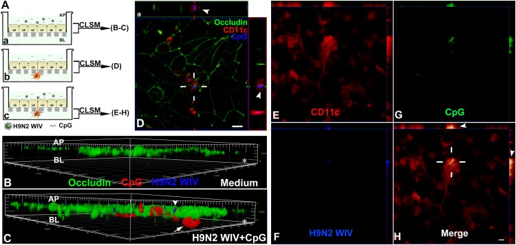
FIG 8.
Uptake of CpGs by both ECs and TEDs. (A) Schematic depicting the Calu-3 monolayer, where DyLight 405-labeled H9N2 WIV plus Alexa Fluor 594-CpGs were seeded on the apical side of the ECs for 1 h (a), and the in vitro DC/EC coculture system, where Alexa Fluor 594-CpGs alone (b) or plus DyLight 405-labeled H9N2 WIV (c) were incubated on the apical side of the Calu-3 monolayer for 1 h. The filters were processed for CLSM. AP, apical side; BL, basolateral side. (B and C) Three-dimensional rendering of representative images obtained using Imaris 7.2 software. (C) Viruses (blue, arrowhead) were only on the apical side of ECs, whereas CpGs (red, arrow) could enter into the ECs. (D) In views taken between the apical side and the filter, cross-sectional images show that TEDs (red [CD11c], arrowheads) crossed the TJs of ECs (occludin, green) and captured the CpGs (blue). (E to H) In views taken between the filter and the basolateral side, cross-sectional images show that both H9N2 WIV (blue) and CpGs (green) existed within submucosal DCs (red [CD11c], arrowheads). The results shown are from a representative experiment out of three. Asterisks indicate the filter. (D to H) Bars: 10 μm.

