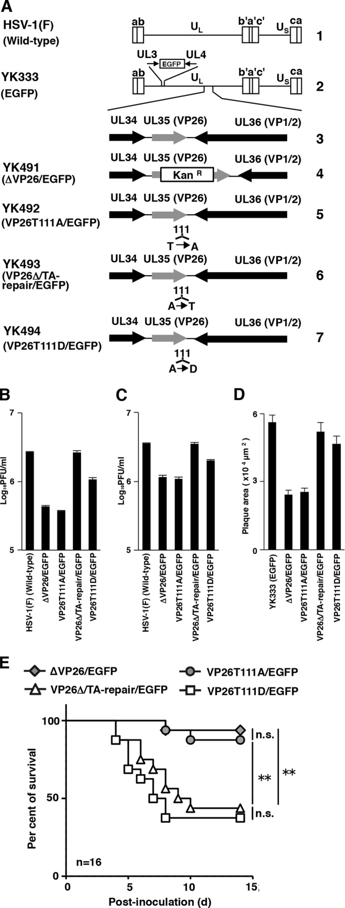
FIG 1.

(A) Schematic diagrams of the genome structure of the wild-type and recombinant HSV-1 viruses used in this study. Line 1, the wild-type HSV-1(F) genome; line 2, the wild-type HSV-1(YK333) genome carrying an expression cassette for EGFP in the intergenic region between UL3 and UL4; line 3, the domain carrying the UL34, UL35(VP26), and UL36(VP1/2) open reading frames; lines 4 to 7, genomes of recombinant viruses with a mutation in the UL35(VP26) gene. (B and C) Effects of mutations in VP26 on progeny virus yield in SK-N-SH cells. SK-N-SH cells were infected with each of the indicated viruses at an MOI of 0.01 (B) or MOI of 5 (C). Total virus from the cell culture supernatants and infected cells was harvested at 48 h postinfection (B) or 24 h postinfection (C) and assayed on Vero cells. Each value is the mean ± standard error of triplicate experiments. Data are representative of three independent experiments. (D) Effect of mutations in VP26 on virus plaque diameter. SK-N-SH cells were infected with each of the indicated viruses at an MOI of 0.00001 under plaque assay conditions. The areas of 25 single plaques for each of the indicated viruses were determined 48 h postinfection. Each value is the mean ± standard error of the measured plaque sizes. Data are representative of three independent experiments. (E) Effect of mutations in VP26 on mortality of mice following intracranial inoculation. Sixteen 3-week-old female ICR mice were inoculated with 500 PFU of the indicated viruses intracranially, and survival was monitored for 14 days. Asterisks indicate significant differences: **, P < 0.0083 (0.05/6) by the log-rank test with Bonferroni adjustment for the four comparison analyses; n.s., not significant.
