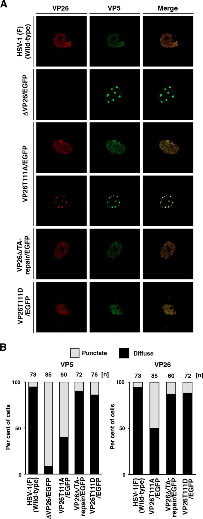
FIG 3.

Effects of mutations in VP26 on the subcellular localization of VP26 and VP5 in SK-N-SH cells. (A) SK-N-SH cells were infected with each of the indicated viruses at an MOI of 5, fixed at 12 h postinfection, permeabilized, stained with antibody to VP26 (red) and VP5 (far red, pseudocolored in green), and examined by confocal microscopy. (B) Quantitation of localization patterns of VP26 and VP5 in the nucleus of the infected SK-N-SH cells shown in panel A. The subcellular localizations of VP26 and VP5, classified into “diffuse” and “punctate,” and frequencies of their localization in infected cells for each of the indicated viruses. The numbers above the columns describe the number of cell samples analyzed. Each data point is representative of the results from three independent experiments.
