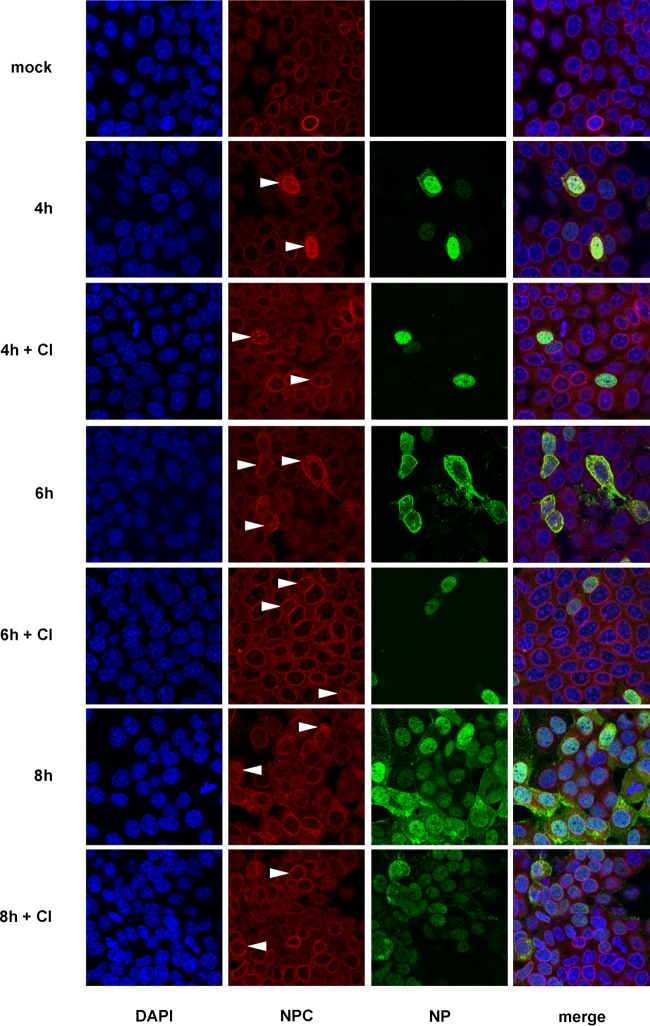
FIG 4.
Nup153 distribution in IV-infected cells. FPV-infected MDCK-II cells (MOI = 1) treated with or without caspase 3/7 inhibitor (CI) were stained for DNA (blue) and for viral NP (green) and with an anti-NPC protein antibody, Mab414, that recognizes FXFG repeats conserved in various nucleoporins (red). Spatial distribution of different Nups at the nuclear envelope was analyzed at 4, 6, and 8 h p.i. by confocal laser scanning microscopy. Partial or (nearly) complete delocalization of Nup in infected cells is indicated by arrowheads.

