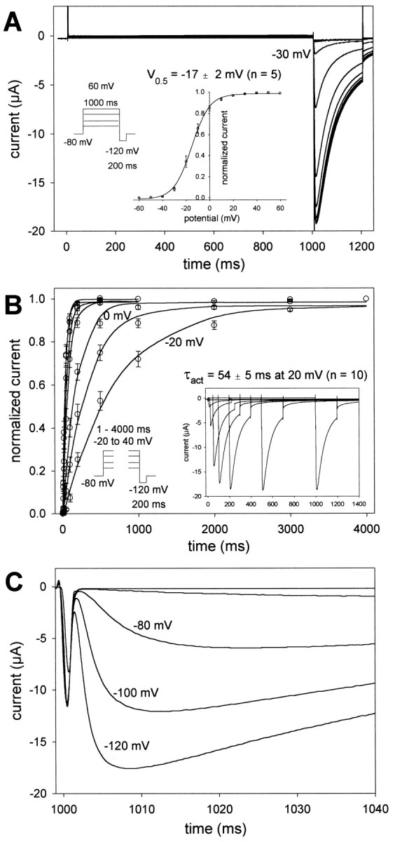
Fig. 2.

Voltage and time dependence of EXP-2 activation and recovery from inactivation. A, Oocytes were held at −80 mV, and 1 sec test pulses to potentials from −60 to 60 mV were applied in 10 mV increments, followed by 200 msec pulses to −120 mV. The first inward tail current could be detected at −30 mV, indicating the onset of activation of EXP-2 channels. Inset,g–V curve with a midpoint of activation V0.5 = − 17 ± 2 mV and slopek = 6.5 ± 0.3 mV per e-fold change in conductance (n = 5 oocytes). B,Oocytes were held at −80 mV, and test pulses to potentials from −20 to 40 mV were applied lasting from 1 to 4000 msec. The time dependence of EXP-2 activation was determined from the peak tail currents during the subsequent pulses to −120 mV. Inset, Actual recording at 20 mV. In A and B, oocytes were bathed in 100 mm KCl in frog Ringer's solution, and current traces were not leak-subtracted. C, EXP-2 channels were activated by 1 sec prepulses to 20 mV. Two hundred millisecond test pulses to different potentials from −120 to −60 mV were applied to initiate recovery from inactivation. Recovery from inactivation is faster at more negative potentials, indicating its steep voltage dependence.
