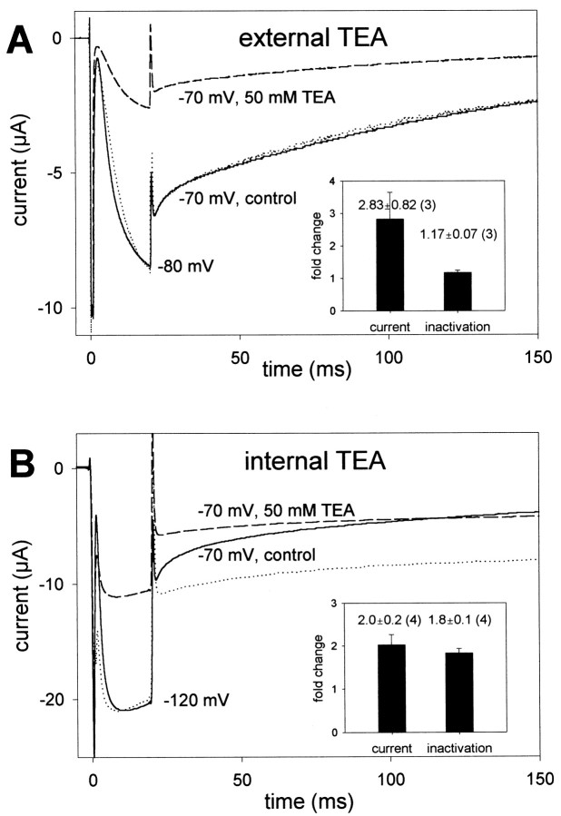
Fig. 6.
Effect of external and internal TEA on inactivation. Fully activated and inactivated EXP-2 channels were allowed to recover from inactivation for 20 msec at −80 or −120 mV, followed by a test pulse to −70 mV to determine the time course of inactivation. A, External application of 50 mm TEA reduces the current amplitude after recovery from inactivation to 31% of control without changing the kinetics of current decay. The dotted line represents the current trace in the presence of TEA scaled to match the control current at the end of recovery from inactivation. Inset, Nearly threefold reduction of current (to 41 ± 10% of control, mean ± SEM; n = 3) without changing the inactivation time constant. B, Internal application of ∼50 mm TEA reduces the current to 53% of control and slows inactivation. The dotted line represents the scaled current trace in presence of TEA for direct comparison with the control trace. Inset, Twofold current reduction and a twofold increase in the inactivation time constant (mean ± SEM;n = 4).

