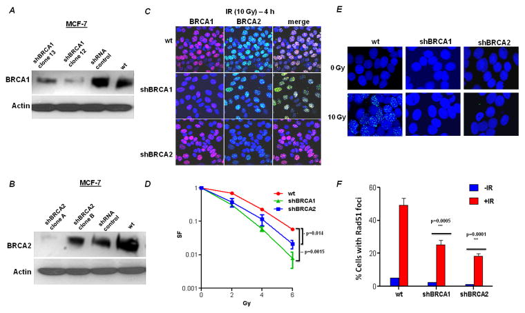
Figure 1. Depletion of BRCA1 and BRCA2 by shRNA in MCF-7 cells.
MCF-7 cells were stably transfected with empty vector (pKLO.1) or with short hairpin RNA constructs against BRCA1 or BRCA2. The efficacy of the shRNA constructs in depleting BRCA1 (A) and BRCA2 (B) was determined by immunoblotting (actin served as loading control) and confirmed by immunofluorescence. (C) Confocal microscopic images of cell clones using immunofluorescent staining for BRCA1 and BRCA2. Wild type (empty vector) and BRCA1 or BRCA2 deficient MCF-7 cells were irradiated (10 Gy) and 4h later, the cells were fixed and processed for BRCA1 (red) or BRCA2 (green) immunofluorescence. The nuclei were identified by DAPI staining (blue). (D) Relative survival of BRCA1- or BRCA2-deficient cells after exposure to IR. Data are the mean and SE of the mean from three independent experiments. The survival of BRCA1- and BRCA2- deficient cells relative to wild type MCF-7 at 6 Gy was compared for statistical significance. (E) Formation of Rad51 foci (green) in response to DNA damage induced by IR. Nuclei were stained with DAPI. Representative nuclei are displayed from either untreated or irradiated cells. Quantification of the percentage of foci positive cells is shown (mean and SE of the mean from three independent experiments).

