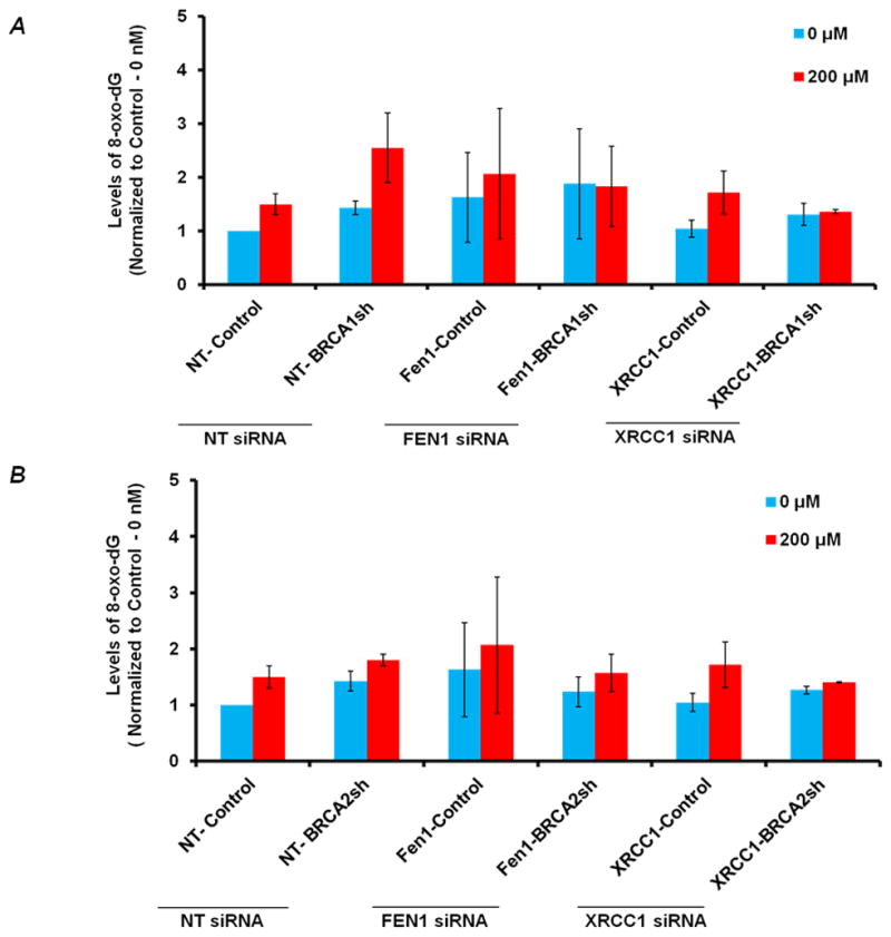
Figure 3. A: Levels of 8-oxo-dG in BRCA1-depleted and FEN1-BRCA1 or XRCC1-BRCA1 co-depleted cells.<.

br>The relative levels of 8-oxo-dG (Mean Fluorescence Intensity, MFI –normalized to 0-nM of control cells) in BRCA1-depleted, FEN1-BRCA1 and XRCC1-BRCA1 co-depleted cells respectively before and after H2O2 treatment. Cells were nucleofected with FEN1 and XRCC1 siRNA respectively and 48 hrs after transfection treated with 200μM of H2O2 for 1 hr. Subsequently, harvested cells were subjected to 8-oxo-dG analysis by flow cytometry. 8-oxodG was determined by using primary antibody against 8-oxodG (Trevigen) and Alexa fluor 488-Rabbit-anti-Mouse secondary antibody IgG (H+L) (Molecular Probes®) using FACS (LSRII). Results are from three independent experiments and are expressed as the mean ± SE. To reduce the basal levels of oxidative DNA damage cells were incubated in 3% Oxygen incubator. B: Levels of 8-oxo-dG in BRCA2-depleted and FEN1-BRCA2 or XRCC1-BRCA2 co-depleted cells: Relative levels of 8-oxo-dG (Mean Fluorescence Intensity, MFI – normalized to 0-nM of control cells) in BRCA2-depleted, FEN1-BRCA2 and XRCC1-BRCA2 co-depleted cells respectively before and after H2O2 treatment as in A.
