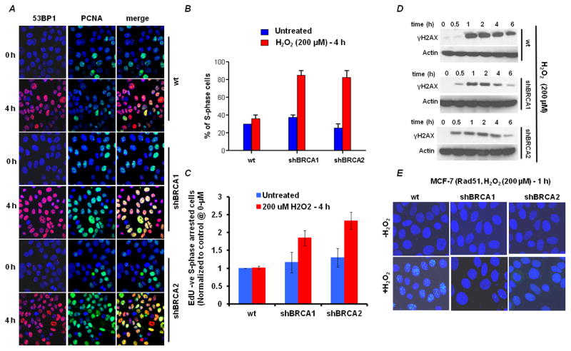
Figure 4. BRCA-deficient cells accumulate in S phase due to oxidative stress-induced double-strand breaks.
(A) Cells were treated with 200μM H2O2 for 4h and the immunofluorescence staining pattern was imaged. Double-strand breaks are shown by 53BP1 (red); S-phase cells are shown by the presence of PCNA in nuclear foci (green). (B) Quantification of the percentage of PCNA foci-positive cells from A. Data shown are the mean and SE from three independent experiments. (C) Quantification of S phase arrested BRCA deficient cells. Cells were treated with 200μM H2O2 for 4h and flow-cytometric analysis of S phase arrested cells after exposure to H2O2 treatment (EdU negative – propidium iodide S phase DNA content) showing reduced incorporation of EdU were measured (additional data in Fig S3). Flow cytometric data is normalized to untreated control cells. Data shown are the mean and SE from three independent experiments. (D) Representative Immunoblots of γH2AX accumulation in BRCA1, BRCA2 deficient and control cells. Actin was used as loading control. Cell lines were treated with 200μM H2O2 for shown time points and were harvested for immunoblotting. (E) Formation of Rad51 foci (green) 4h after treatment with 200μM H2O2 for 1h.

