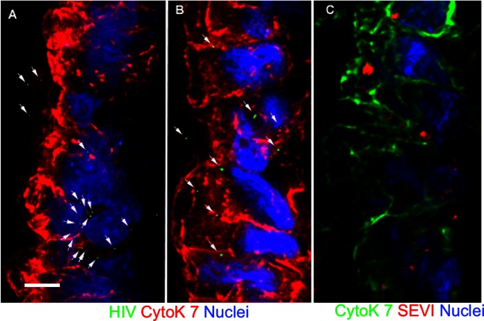
FIG 4.
Untreated and SEVI-treated HIV-1 particles enter the epithelium similarly in human endocervical explants. SEVI fibrils were preincubated with PA–HIV-1. (A and B) Either PA–HIV-1 alone (A) or PA–HIV-1/SEVI complexes (B) were incubated with endocervical explants. (C) Physical appearance of SEVI within the simple columnar epithelium of the endocervix. Arrows in panels A and B highlight HIV-1 within the epithelium. The simple columnar epithelium is visualized by cytokeratin 7 staining and nuclear Hoechst staining. For each panel, the lumen is on the left side of the image. Images were taken at a ×100 magnification. Bars = 5 μm.

